

Ultrasound Based Spinal Navigation
Projektleiter:
Projektbearbeiter:
M.Sc. Sathish Balakrishnan,
M.Sc. Nazila Esmaeili,
M.Sc. Jens Ziegle,
Dr. Alfredo Illanes,
M.Sc. Elmer Jeto Gomes Ataide,
Dr.-Ing. Axel Boese
Finanzierung:
Industrie;
Forschergruppen:
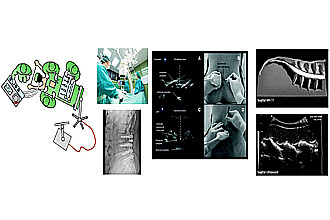
Intra-operative Navigation during a spinal surgery is conventionally carried out using X-Ray images from a C-ARM device. Additionally invasive markers are attached to the patients bone to have a fixed reference coordinate system for the C-ARM, which can be used for fusing the X-Ray images with a pre-operative CT or MRI. Further this fusion information is used to intra-operatively locate the spinal bones for drilling. Even though this procedure is the state of art, it is inhibited by the X-ray radiation exposure to both surgeon and the patient, and using a C-ARM occupies a lot of space which inhibits the mobility inside an operating room.
Ultrasound(US) is the cheapest, non-invasive and easily portable imaging modality compared to other conventional imaging modalities like MRI and CT. These advantages of US motivated us to replace C-ARM X-ray images with US images for navigation in a spinal surgery. However suboptimal quality of real-time US images and speckle noise patterns make bone detection in US images harder compared to X-Ray images. In order to achieve this goal, we propose a fast, image based bone segmentation and 2D-3D registration framework that operates with a tracked 2D US images and preoperative CT or MRI 3D volumes.
Our approach clearly reduces the intra-operative radiation exposure and huge space occupied by C-ARM inside the operating room.
Kooperationen im Projekt
Kontakt

Prof. Dr. Michael Friebe
Otto-von-Guericke-Universität Magdeburg
Leipziger Str. 44
39120
Magdeburg
Tel.:+49 391 6715750
weitere Projekte
Die Daten werden geladen ...